

Optos California Retinal Imaging
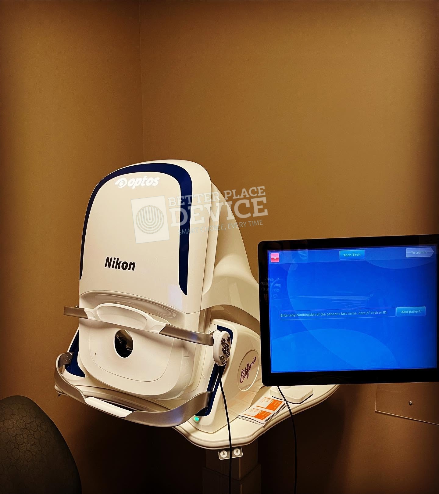
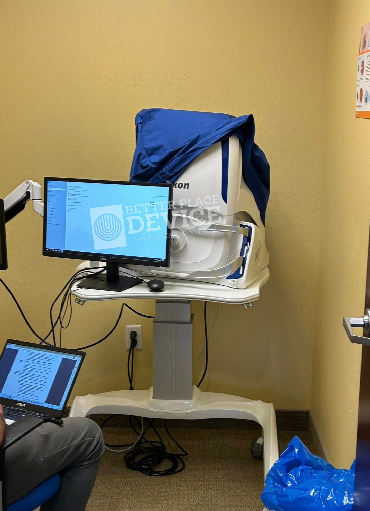
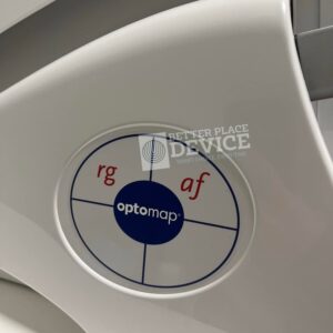
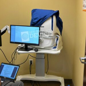
Optos California Retinal Imaging is the multimodal ultra-widefield platform from Optos — single-capture 200° Optomap retinal imaging through pupils as small as 2 mm, plus green/blue autofluorescence, fluorescein angiography, and ICG angiography in one session without dilation. This used optos california ships ready for full retina-clinic and screening workflows. The California’s headline advantage is single-capture coverage of the peripheral retina that conventional 45° fundus cameras (Topcon NW-series, Zeiss Visucam) require multiple shots and montaging to approximate — making it the standard of care for diabetic retinopathy screening, peripheral retinal tear detection, and uveitis workups where peripheral pathology is clinically important. Optomap output integrates directly with EMR via standardized DICOM-style export.
Technical Specifications:
Image Modalities: optomap color and optomap plus (red and green laser):
Color composite view
Green laser view
Red laser view
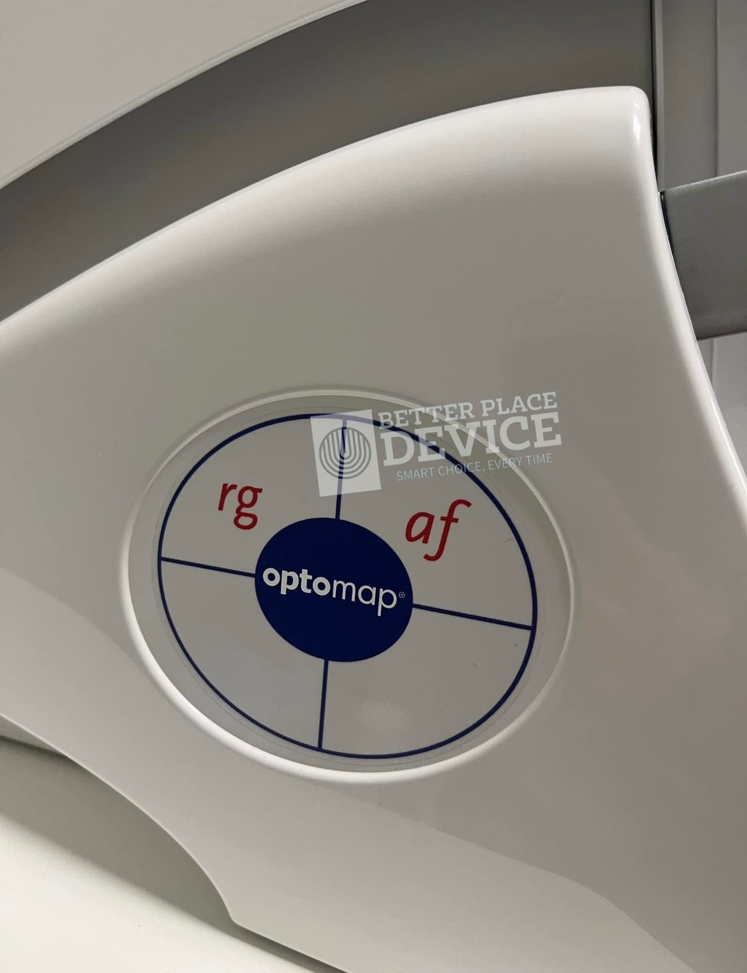
optomap af (green laser): autofluorescence
Resolution: optomap color: 20 μm, optomap plus, af : 14 μm
Wavelengths: Red laser: 635 nm, Green laser: 532 nm
Exposure Time: Less than 0.4 seconds
Foot Print: Width: 550 mm / 22 inches
Depth: 550 mm / 22 inches including chinrest
Height: 608-632 mm / 24-25 inches
Weight: 75 lbs
Communication Protocol: DICOM Compatible
With California, Optos has incorporated new hardware and software technology enabling practitioners to see more, discover more and effectively treat more ocular pathology thus promoting patient health. We are committed to further strengthening our clinical evidence while demonstrating the importance of imaging the entire retina.
We have created three versions of California to be customizable for use in multiple eyecare settings. California includes a new UWF optomap imaging modality; Indocyanine Green angiography (icg) while retaining –
Composite color
Red-free
Auto fluorescence (af)
Fluorescein angiography (fa)
Images are now presented in ProView which displays optomap in a consistent geometry that accurately represents anatomical features in the retina. Further, ProView enables automatic image registration for disease tracking over time, and inter-modality image comparison. New proprietary optical hardware optimizes and maintains resolution of the optomap images throughout the scan of the retina resulting in more clarity in the far periphery.
Be the first to review “Optos California Retinal Imaging”
Related products
Ophthalmology Equipment
Ophthalmology Equipment
Ophthalmology Equipment
Ophthalmology Equipment
Reviews
There are no reviews yet.